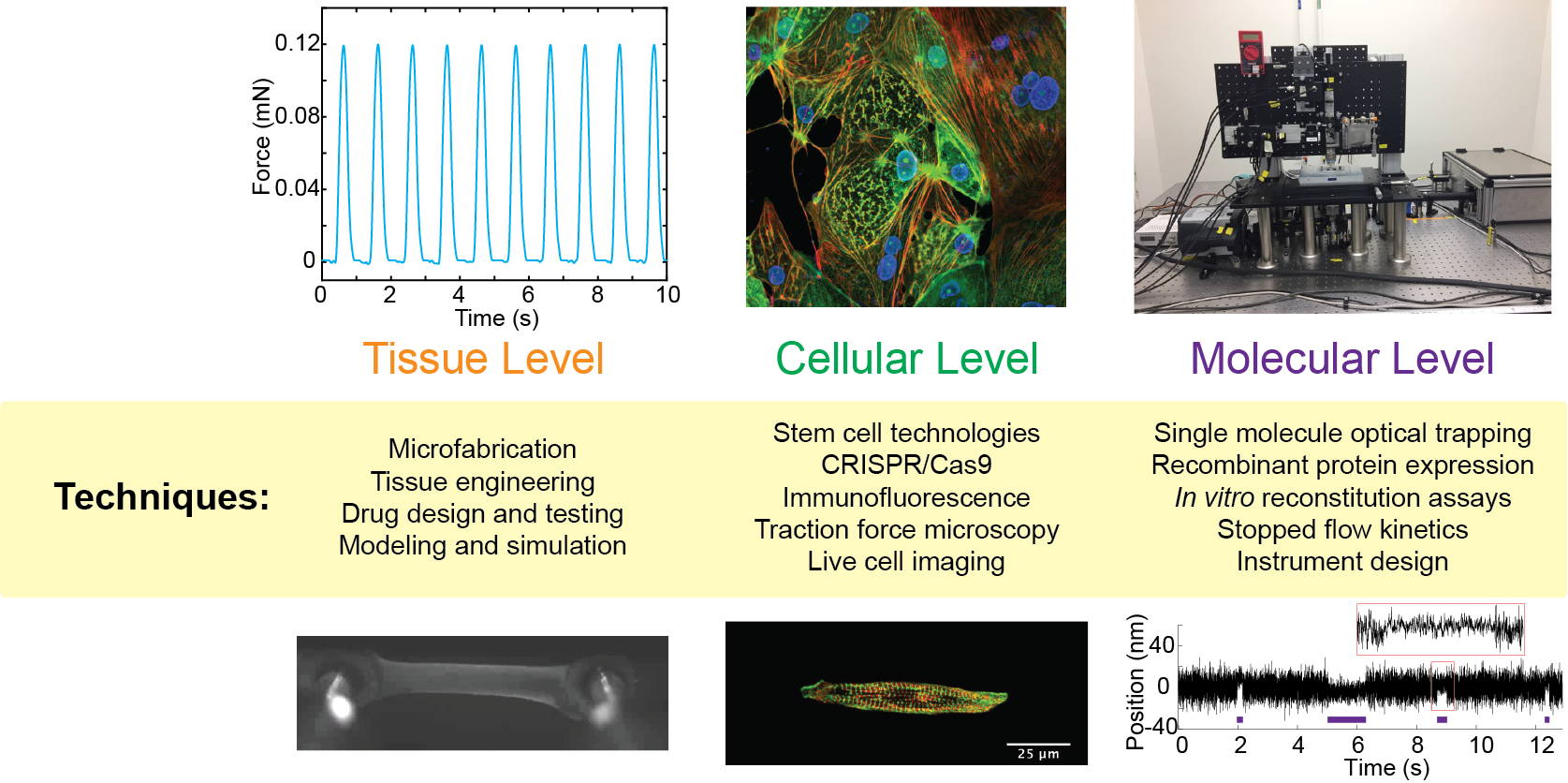
The Greenberg Lab takes an interdisciplinary approach to understanding molecular motors over multiple levels of organization. Techniques used by the Greenberg Lab include:
Single Molecule Optical Trapping
We use optical trapping to examine the contractile properties of single myosin molecules. Our home-built optical trap enables us to measure nanometer movements and piconewton forces with high spatial and temporal resolution. Moreover, our system enables us to simultaneously measure single molecule fluorescence using total internal reflectance microscopy. We are using this system to reconstitute muscle contraction at the molecular level.
Stopped Flow Rapid Kinetics and Biochemical Assays
We use biochemical and biophysical techniques to examine the interactions between motor proteins and binding partners. Our stopped-flow apparatus allows us to measure the rates of very fast biochemical reactions. Using this technique, it is possible to dissect the individual steps of the myosin ATPase cycle. We also use other biochemical and biophysical techniques including fluorimetry, sedimentation assays, and chromatography.
Cell Culture and Stem Cell Engineering
Our lab uses stem cell technologies to model heart disease. We use the CRISPR/Cas9 system to introduce disease-causing mutations into human stem cells and then we differentiate these cells to cardiomyocytes. We also culture mammalian cells for use in engineered tissues. Click on the image above to see the beating cardiac tissue.
Traction Force Microscopy
To examine the contractile properties of stem-cell derived cardiomyocytes, we use traction force microscopy. We use microfabrication techniques to pattern extracellular matrix proteins on to a polyacrylamide hydrogel with defined mechanical properties. As the cell beats, beads embedded in the gel are displaced, allowing for the calculation of the cellular force of contraction. Click on the image above to see a beating cardiomyocyte on the hydrogel.