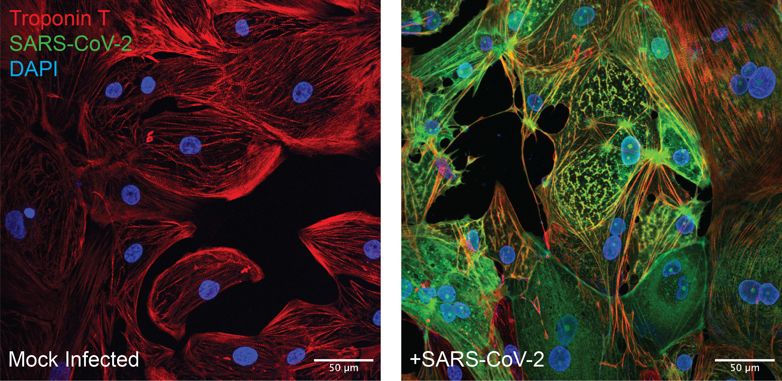
 Human stem cell derived cardiomyocytes infected with SARS-CoV-2 show sarcomere disassembly.
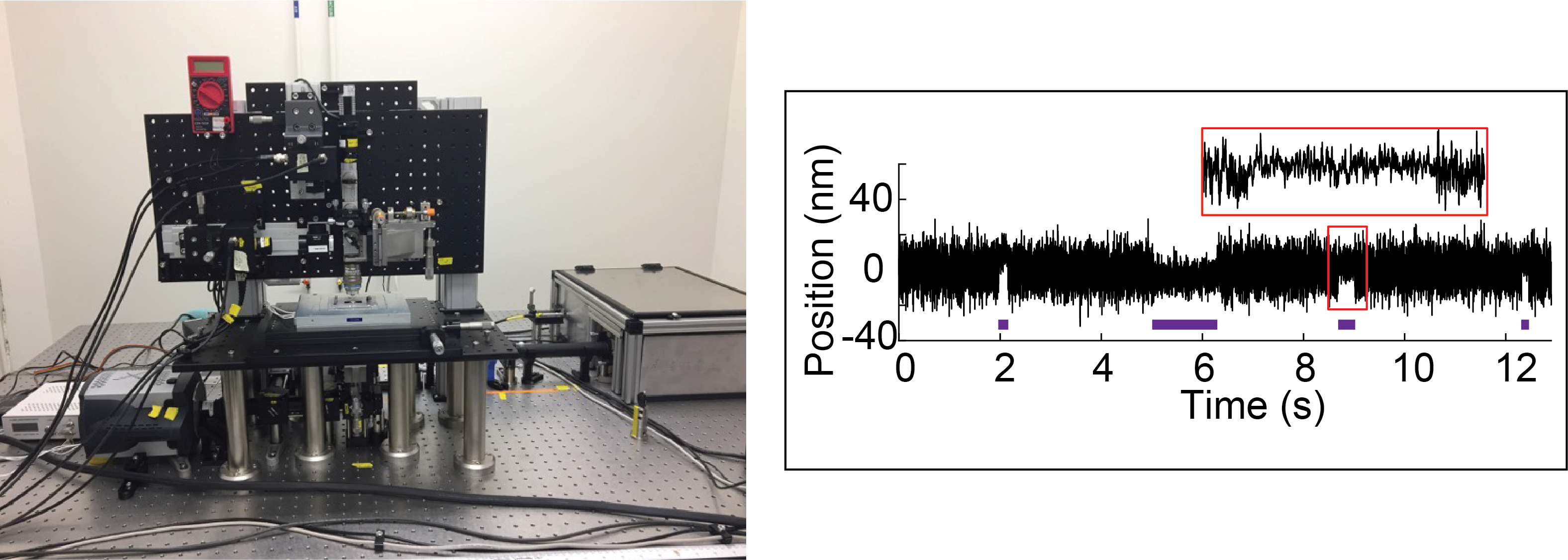
(Left) Our custom-built dual beam optical trapping microscope. (Right) Data trace showing single molecule interactions between cardiac myosin and actin. Binding interactions are identified by purple lines.
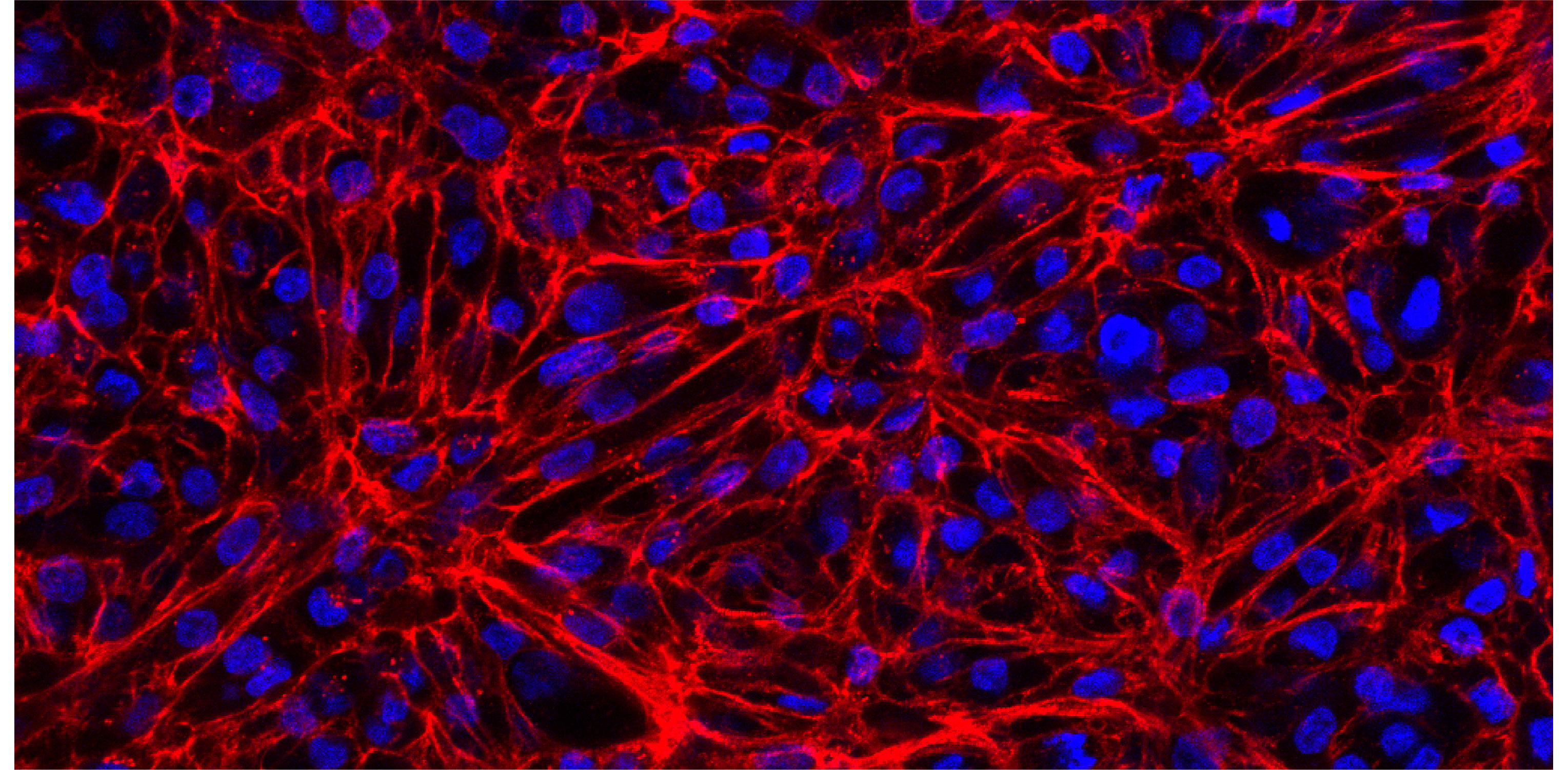 Confocal image of a human engineered heart tissue stained for the sarcomeric marker troponin T (red) and DAPI (blue).
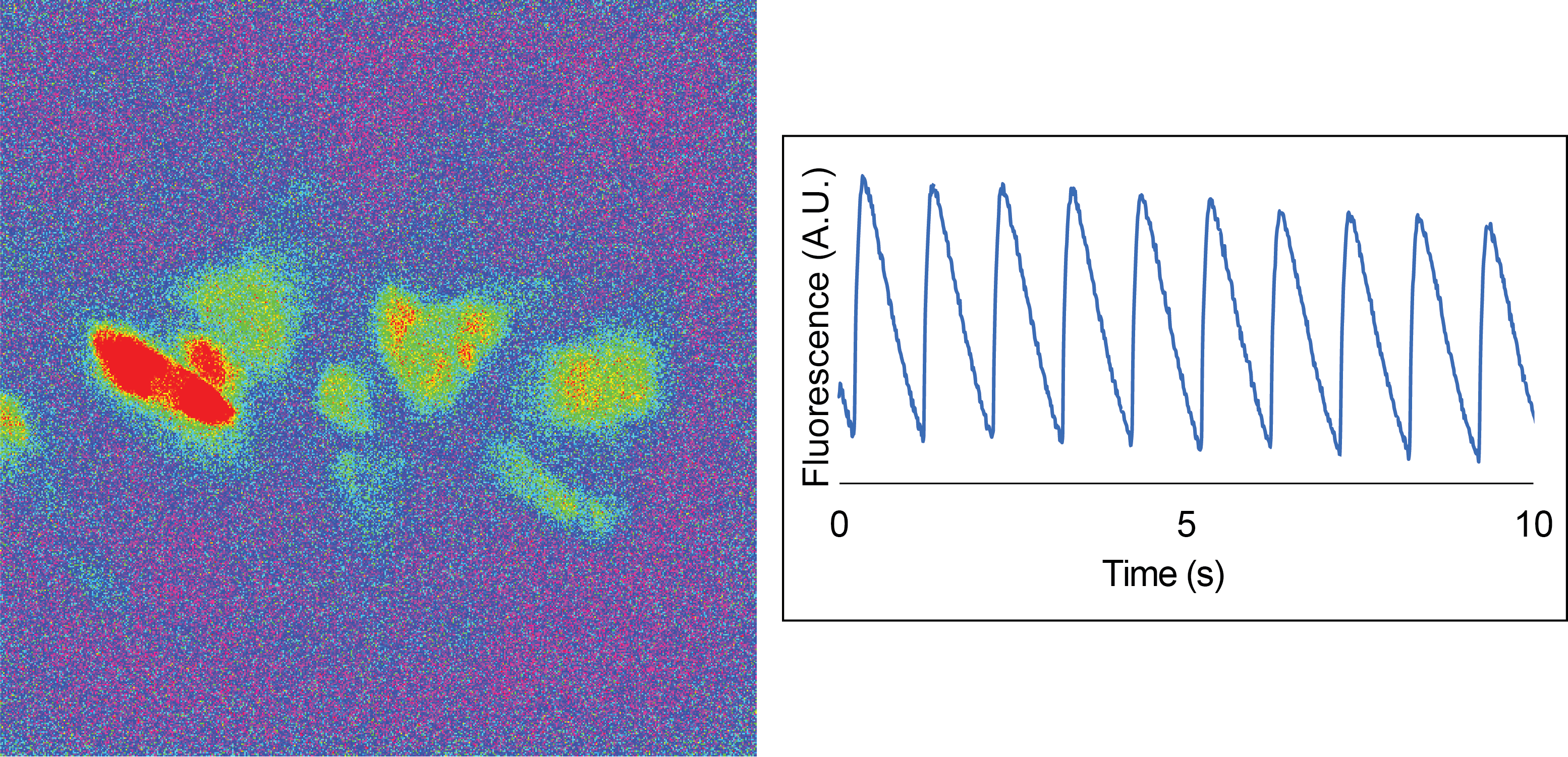
Human stem cell derived cardiomyocytes display calcium transients when electrically stimulated. (Left) Image showing calcium-induced fluorescence. (Right) Data trace showing calcium transients. The fluorescence is plotted as a function of time. Cells are stimulated at 1 Hz.